Home » Without Label » Anatomy Of Back Of Neck - Anatomy Of The Back Spine And Back Muscles Kenhub : Corenman earned academic appointments as clinical assistant.
Anatomy Of Back Of Neck - Anatomy Of The Back Spine And Back Muscles Kenhub : Corenman earned academic appointments as clinical assistant.
Anatomy Of Back Of Neck - Anatomy Of The Back Spine And Back Muscles Kenhub : Corenman earned academic appointments as clinical assistant.. The splenius muscles originate at the midline and run laterally and superiorly to their insertions. It serves as the connecting point between the head and the trunk. It also covers some common conditions and injuries that can affect the. Neck muscles help support the cervical spine and contribute to movements of the head, neck, upper back, and posterior longitudinal ligament (pll). Surface anatomy and surface markings.
Head and neck anatomy is important when considering pathology affecting the same area. The back muscles stabilize and move the vertebral column the splenius muscles originate at the midline and run laterally and superiorly to their insertions. Develop students understanding of the ways in which structure and function of muscle and joints. Resists back hyperextension, c1 to sacrum resists hyperflexion of the back, helps prevent herniation, c2… Trained as both a medical doctor and doctor of chiropractic, dr.
Anatomy Of The Spine Blog Back Pain Neck Pain Newark New Jersey from www.suborthonj.com Our neck is where we find the seven cervical vertebrae, with c7 (the seventh cervical vertebra) meeting t1 (the first thoracic vertebra) at the base of the neck. Neck, in land vertebrates, the portion of the body joining the head to the shoulders and chest. This article describes the anatomy of the head and neck of the human body, including the brain, bones, muscles, blood vessels, nerves, glands, nose, mouth, teeth, tongue, and throat. Home/anatomy/anatomy of the cervical spine. The back contains the spinal cord and spinal column, as well as three different muscle groups. Trained as both a medical doctor and doctor of chiropractic, dr. The pll starts at c2 and goes down the back of the vertebral bodies and intervertebral discs. The word neck comes from a latin word which means cervical.
It serves as the connecting point between the head and the trunk.
The cervical spine supports the weight and movement of your head and protects the nerves exiting your brain. In radiology, the 'head and neck' refers to all the anatomical structures in this region excluding the central nervous system, that is, the brain and spinal co. The neck is the area between the skull base and the clavicles. 3d video tutorials and interactive modules on the anatomy of the back including anatomy of the musculature, vertebral column, joints and ligaments. Back view of anatomy of male back pain in blue. A collection of anatomy notes covering the key anatomy concepts that medical students need to learn. Despite being a relatively small region, it contains a range of important anatomical features. Watch cervical muscle anatomy animation. « back show on map ». By david terfera, shereen jegtvig. Our neck is where we find the seven cervical vertebrae, with c7 (the seventh cervical vertebra) meeting t1 (the first thoracic vertebra) at the base of the neck. Your neck is like no other part of the vertebral spinal column and enables your head and neck a wide range of motion. Trained as both a medical doctor and doctor of chiropractic, dr.
Foundational anatomy provides medical students with the necessary background in anatomy for success in clerkships. The cervical spine supports the weight and movement of your head and pro. In the neck, the platysma when contracted throws the skin into oblique ridges parallel with the fasciculi of the muscle. Back view of anatomy of male back pain in blue. The head rests on the top part of the vertebral column, with the skull joining at c1.
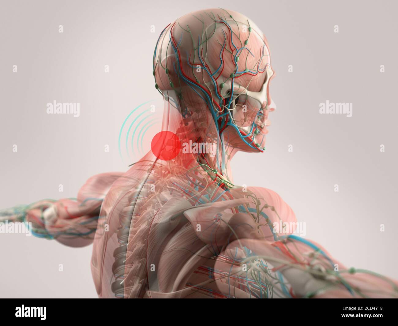
Human Anatomy Neck Pain Head Shoulders And Back Stock Photo Alamy from c8.alamy.com All of the anatomical structures of the face with labels on 150 axial and coronal slices from a scan: The posterior muscles of the neck are primarily concerned with head movements, like extension. Clinically, surface anatomy is used to split the neck into anterior and posterior triangles which provide clues as to the location of specific structures. Muscle head anatomy vocal organ diagram female neck anatomy neck wireframe head neck human anatomy head artery anatomy face pharynx vector neck degree head anatomy 3d. It is made up of bones discs muscles ligaments nerves and tendons. From the sides and the back of the neck, the splenius capitis inserts onto the head region, and the splenius cervicis extends onto the cervical region. Neck, in land vertebrates, the portion of the body joining the head to the shoulders and chest. By david terfera, shereen jegtvig.
Demonstrate sound knowledge of the surface/living and radiological anatomy of the head, neck and.
A coronal and axial contrast enhanced multidetector computed tomography imaging of the head and neck was performed on a healthy subject. Our neck is where we find the seven cervical vertebrae, with c7 (the seventh cervical vertebra) meeting t1 (the first thoracic vertebra) at the base of the neck. It also covers some common conditions and injuries that can affect the. Head and neck anatomy is important when considering pathology affecting the same area. Some important structures contained in or passing through the neck include the seven cervical vertebrae and enclosed spinal cord, the jugular veins and carotid arteries, part of the esophagus, the larynx. The structure is, of course, an important part of the conversation. In the neck, the platysma when contracted throws the skin into oblique ridges parallel with the fasciculi of the muscle. Many conditions and injuries can affect the back. Learn about the various causes of back pain, including different kinds of arthritis. The cervical spine supports the weight and movement of your head and protects the nerves exiting your brain. By understanding the anatomy of the neck and how each structure works, it's easier to understand the sources of neck pain. It is made up of bones discs muscles ligaments nerves and tendons. The clinical anatomy of the head, neck sternocleidomastoid muscle (main muscle in the front of the neck).
The neck is considered to be a very important part of the body as it serves to support the head. The arteries that ultimately supply the head and neck originate from the subclavian and common carotid arteries. Additionally, the joints in the back of the cervical vertebrae (facets) are shaped to allow movement: The physicians originally studying human anatomy thought the skull looked like an helmet. Watch cervical muscle anatomy animation.
Spine Structure Function Parts Segments Spine Problems Spine Health from www.clevelandclinic.org The cervical spine supports the weight and movement of your head and pro. The arteries that ultimately supply the head and neck originate from the subclavian and common carotid arteries. All of the anatomical structures of the face with labels on 150 axial and coronal slices from a scan: The cervical spine supports the weight and movement of your head and protects the nerves exiting your brain. The pll starts at c2 and goes down the back of the vertebral bodies and intervertebral discs. It is made up of bones discs muscles ligaments nerves and tendons. By david terfera, shereen jegtvig. Want to learn more about it?
Your neck is like no other part of the vertebral spinal column and enables your head and neck a wide range of motion.
From the sides and the back of the neck, the. In radiology, the 'head and neck' refers to all the anatomical structures in this region excluding the central nervous system, that is, the brain and spinal co. Home/anatomy/anatomy of the cervical spine. Want to learn more about it? Our neck is where we find the seven cervical vertebrae, with c7 (the seventh cervical vertebra) meeting t1 (the first thoracic vertebra) at the base of the neck. Head and neck anatomy is important when considering pathology affecting the same area. From the sides and the back of the neck, the splenius capitis inserts onto the head region, and the splenius cervicis extends onto the cervical region. The neck muscles, including the sternocleidomastoid and the trapezius, are responsible for the gross motor movement in the muscular system of the head and neck. The back muscles stabilize and move the vertebral column the splenius muscles originate at the midline and run laterally and superiorly to their insertions. The splenius muscles originate at the midline and run laterally and superiorly to their insertions. Neck, in land vertebrates, the portion of the body joining the head to the shoulders and chest. Neck muscles help support the cervical spine and contribute to movements of the head, neck, upper back, and posterior longitudinal ligament (pll). The arteries that ultimately supply the head and neck originate from the subclavian and common carotid arteries.